
Published on: Aug 30, 2023
Last updated: October 5, 2026
Next review: October 2027, or sooner if major ophthalmology or stroke guidance changes.
Written and source checked by: Adel Galal, Founder and Lead Writer at NextFitLife
Medical review status: This article has not been medically reviewed by an ophthalmologist, retina specialist, neurologist, cardiologist, or other licensed healthcare professional.
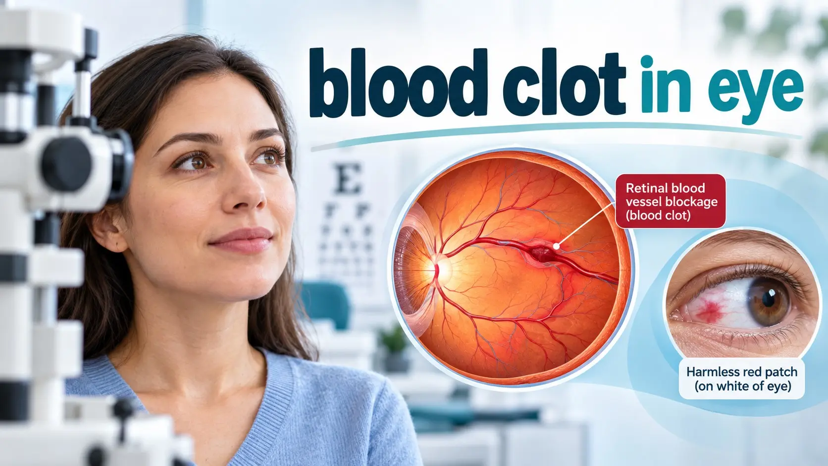
A blood clot in eye can mean a blockage in a retinal artery or vein, but the phrase is also used incorrectly for a bright red patch on the white of the eye. Sudden painless loss of vision in one eye can signal a retinal artery occlusion, or eye stroke, and needs emergency assessment.
The first job is therefore not to ask, “How do I dissolve the clot?”
It is to identify what kind of eye problem you actually have.
Medical Notice: This content is for general educational purposes and does not replace professional medical advice, diagnosis, or treatment. Consult an appropriately qualified healthcare professional for personal medical concerns.
Sudden vision loss, a new missing area of vision, severe eye pain, eye symptoms with facial weakness or speech trouble, or sudden vision loss in an adult over 50 with a new headache or jaw pain can require emergency care.
Table of Contents
- What Blood Clot in Eye Really Means
- Retinal artery blockage
- Retinal vein blockage
- Surface bleeding that is not a retinal clot
- 9 Critical Warning Signs
- Sudden painless vision loss
- Sudden blurry vision
- A missing area of vision
- Vision change in one eye
- New visual distortion
- Redness with vision loss
- Eye pain with new visual symptoms
- Stroke symptoms
- Headache and jaw pain after age 50
- Main Causes and Risk Factors
- How Eye Doctors Diagnose the Problem
- Treatment
- What Not to Do
- Prevention
- Frequently Asked Questions
What does “blood clot in the eye” actually mean?
The phrase is not one precise medical diagnosis.
People use it to describe several very different conditions.
The most important ones are:
- Retinal artery occlusion, where blood flow into part or all of the retina is suddenly blocked
- Retinal vein occlusion, where blood has trouble draining out of the retina
- Subconjunctival hemorrhage, where a tiny surface blood vessel breaks and leaves a red patch on the white of the eye
These do not have the same urgency, cause, or treatment.
That distinction is the foundation of this guide.
For a broader overview of vision care, use the NextFitLife Eye Health Complete Guide.
Is a bright red patch on the white of the eye a blood clot?
Usually, no.
A sudden bright red patch on the white part of the eye is often a subconjunctival hemorrhage.
This happens when a tiny blood vessel under the clear surface tissue of the eye breaks and leaks blood.
It can look dramatic.
It is usually harmless.
Typical features include:
- A sharply visible red patch on the white of the eye
- No change in vision
- No major eye pain
- No pus or eye discharge
Coughing, sneezing, vomiting, straining, rubbing the eye, or a minor bump can trigger it.
High blood pressure, diabetes, some blood thinning medicines, and bleeding disorders can also raise the chance of repeated episodes.
The blood is usually absorbed by the body over days to a few weeks.
A surface hemorrhage is therefore very different from a blocked retinal vessel behind the eye.
What is retinal artery occlusion?
A retinal artery occlusion happens when blood flow through an artery feeding the retina becomes blocked.
The retina is the light sensing tissue at the back of the eye.
It needs constant oxygen rich blood.
A blockage can quickly injure retinal tissue.
Central retinal artery occlusion
Central retinal artery occlusion, or CRAO, blocks the main retinal artery.
The American Heart Association classifies CRAO with retinal infarction as a form of acute ischemic stroke.
That is why you may hear the term eye stroke.
This is a medical emergency.
Branch retinal artery occlusion
A branch retinal artery occlusion affects a smaller retinal artery.
Instead of losing most vision in the eye, a person may lose part of the visual field.
Acute symptomatic retinal artery occlusion still deserves emergency assessment because it can point to wider vascular disease.
1. Sudden painless loss of vision is the biggest warning sign
CRAO classically causes sudden, painless vision loss in one eye.
The change can happen over seconds.
This is very different from slow vision decline over months or years.
Do not:
- Wait for vision to return
- Go to sleep and check again tomorrow
- Try eye drops first
- Massage the eye
- Drive yourself if vision is badly affected
Seek emergency medical assessment.
2. Sudden blurred vision can signal retinal vein occlusion
Retinal vein occlusion blocks the vessel that normally carries blood away from the retina.
This causes blood and fluid to back up.
The retina can then develop bleeding, swelling, and poor oxygen supply.
A central retinal vein occlusion, or CRVO, affects the main retinal vein.
A branch retinal vein occlusion affects a smaller branch.
Blurred vision is one of the most common symptoms.
Vision loss may be mild or severe depending on the area involved.
3. A missing patch of vision can signal a branch artery blockage
Not every retinal artery blockage causes complete blindness in one eye.
A blockage in one branch can remove part of the visual field.
You might notice that:
- One side of vision looks dark
- A section seems missing
- Objects disappear in one area
- Reading suddenly becomes difficult despite one part of vision remaining clear
A new field defect needs urgent eye and medical assessment.
4. A sudden change affecting only one eye is an important clue
Many retinal vessel occlusions affect one eye.
A simple way to notice this is to cover one eye and then the other.
Do this only to describe the symptom.
Do not let self testing delay emergency care.
Sudden one eye vision loss can also come from retinal detachment, optic nerve disease, eye bleeding, or other emergencies.
You cannot safely tell them apart without examination.
5. Distorted or blurred central vision can come from macular edema
A retinal vein occlusion can increase a protein called VEGF inside the eye.
This can make retinal blood vessels leak.
Fluid may collect in the macula, which is responsible for sharp central vision.
This is called macular edema.
You may notice:
- Blurred central vision
- Distortion
- Difficulty reading
- Loss of fine detail
This is one reason retinal vein occlusion treatment often focuses on controlling macular swelling.
6. Redness with vision loss is not a simple broken surface vessel
A harmless subconjunctival hemorrhage should not normally cause loss of vision.
That gives you a useful dividing line.
| What You Notice | Possible Problem | Action |
|---|---|---|
| Bright red patch on white of eye, normal vision, little or no pain | Subconjunctival hemorrhage | Usually not an emergency, but seek care if recurrent, traumatic, painful, or associated with vision change |
| Sudden painless major vision loss in one eye | Retinal artery occlusion or another serious eye problem | Emergency assessment now |
| Sudden blurry or reduced vision in one eye | Retinal vein occlusion or another retinal problem | Prompt eye assessment |
| Flashes, many new floaters, or curtain across vision | Possible retinal tear or detachment | Urgent eye assessment |
| Red eye with severe pain, halos, nausea, or sudden blur | Possible acute glaucoma or other emergency | Emergency care |
7. Eye pain with a new vision change needs urgent evaluation
Most CRAO is painless.
That does not mean painful eye symptoms are safer.
Severe pain plus sudden visual change can point toward other urgent conditions such as acute angle closure glaucoma, inflammation, infection, or serious eye injury.
Do not use pain alone to decide whether vision loss is dangerous.
8. Eye symptoms plus stroke symptoms are an emergency
A retinal artery blockage can share causes with a brain stroke.
Call emergency services if eye symptoms occur with:
- Face drooping
- New weakness or numbness on one side
- Trouble speaking
- Trouble understanding speech
- Sudden severe balance trouble
- Confusion
AHA guidance treats CRAO as an acute ischemic stroke involving the retina.
The person therefore needs more than an eye exam.
Stroke risk and the source of the blockage also need evaluation.
9. Sudden vision loss after age 50 with a new headache can signal giant cell arteritis
This deserves its own section because treatment cannot wait.
Giant cell arteritis is inflammation of medium and large arteries.
It occurs in adults over age 50 and can block blood flow to structures supplying the eye.
Warning signs can include:
- New headache
- Scalp tenderness
- Pain in the jaw while chewing
- Unexplained fever
- Fatigue
- Weight loss
- Sudden vision loss
If an older adult develops sudden vision loss with these symptoms, seek emergency medical care.
Doctors may need urgent blood tests and treatment because the other eye can also be at risk.
What causes retinal artery occlusion?
Many cases are linked with an embolus or other interruption of arterial blood flow.
The material blocking the retinal artery can come from elsewhere in the vascular system.
Possible sources include the carotid arteries or heart.
That is why CRAO triggers a stroke style medical evaluation.
Risk factors include:
- High blood pressure
- High cholesterol
- Diabetes
- Smoking
- Obesity
- Cardiovascular disease
- Older age
- Some clotting disorders
This is also why the old idea of treating an eye clot with hydration or “blood thinning foods” is too simplistic.
What causes retinal vein occlusion?
The exact mechanism depends on which retinal vein is blocked.
In CRVO, blood flow through the central retinal vein becomes blocked or severely reduced.
In branch retinal vein occlusion, a smaller retinal vein becomes obstructed, often around a point where an artery and vein cross.
Major risk factors include:
- High blood pressure
- Diabetes
- Glaucoma
- Hardening of the arteries
- Cardiovascular disease
- Older age
Not everyone with one of these conditions develops retinal vein occlusion.
Risk factors tell us who is more vulnerable.
They do not diagnose the condition.
Does dehydration cause a blood clot in the eye?
I would not use dehydration as a main explanation for retinal artery or vein occlusion in an otherwise healthy person.
Retinal vessel blockages have several possible causes and cannot be explained by a simple claim about water intake.
Normal hydration is good for overall health.
Drinking extra water does not treat a retinal vascular occlusion and does not replace control of blood pressure, diabetes, cholesterol, smoking, glaucoma, or cardiovascular disease.
Do blood thinning foods prevent an eye clot?
No food has been proven to act as a substitute for medical prevention or treatment.
Garlic, ginger, omega 3 foods, herbs, and supplements should not be used as DIY anticoagulant treatment for a retinal vessel blockage.
Some supplements can also increase bleeding risk or interact with prescribed medicines.
Do not start aspirin, an anticoagulant, or a high dose supplement because you are worried about an eye clot unless a qualified clinician recommends it.
For broader nutrition support, see 10 Best Foods for Eye Health and Better Vision.
How is a suspected blood clot in the eye diagnosed?
The tests depend on whether the doctor suspects an artery problem, vein problem, surface hemorrhage, retinal detachment, or another cause of vision loss.
Dilated eye examination
Drops widen the pupil so the ophthalmologist can inspect the retina, optic nerve, and blood vessels.
This can reveal retinal hemorrhages, swelling, pale ischemic retina, abnormal vessels, or other signs.
Optical coherence tomography
OCT creates detailed cross sectional images of the retina.
It is especially useful for finding and monitoring macular edema.
Fluorescein angiography
A dye is injected into a vein and photographs are taken as it travels through retinal blood vessels.
This helps show abnormal filling, leakage, and areas of reduced retinal circulation.
Stroke and cardiovascular testing
Retinal artery occlusion needs systemic evaluation.
Testing can include:
- Blood pressure measurement
- Blood glucose testing
- Cholesterol testing
- Heart rhythm assessment
- Carotid artery imaging
- Heart evaluation
- Brain imaging when appropriate
Blood tests for giant cell arteritis
For adults over 50 with suggestive symptoms, clinicians can order ESR, CRP, and blood counts while arranging urgent treatment and further assessment.
How is retinal artery occlusion treated?
The first treatment is speed.
A symptomatic retinal artery occlusion should be treated as an emergency and assessed at a stroke capable medical center.
AHA guidance says intravenous alteplase, a clot dissolving medicine, may be considered in selected patients with disabling visual loss who meet the normal criteria for systemic alteplase.
This is a specialist emergency decision.
It is not routine treatment for every person with sudden vision loss.
Some endovascular approaches are also being studied or considered in specialized situations, but evidence remains limited.
Do not massage the eye
Older advice sometimes suggested ocular massage to move a retinal artery blockage.
AHA guidance states that eye massage, anterior chamber paracentesis, and hemodilution have not been proven effective and may cause harm.
Do not delay emergency treatment while trying these methods.
How is retinal vein occlusion treated?
Retinal vein occlusion is treated differently.
There is no simple procedure that removes the clot and returns the retinal vein to normal.
Treatment focuses on the problems caused by the blockage.
Anti VEGF injections
Anti VEGF injections are commonly used to reduce macular edema from CRVO.
They can improve or stabilize vision in many patients.
More than one injection is commonly needed.
Steroid treatment
Intraocular steroid medicine can help reduce retinal swelling in selected patients.
Laser treatment
Laser treatment may be used when severe retinal ischemia causes abnormal new blood vessels.
These vessels can cause bleeding and dangerous increases in eye pressure.
Can a retinal vein occlusion cause glaucoma?
Yes.
A severe ischemic retinal vein occlusion can trigger growth of abnormal blood vessels.
If these vessels grow around the drainage angle of the eye, eye pressure can rise sharply.
This is called neovascular glaucoma.
It is one reason follow up after CRVO matters even if the first visit is already complete.
Can retinal vein occlusion cause permanent vision loss?
Yes.
The outcome depends on how severe the blockage is, how much retinal ischemia occurs, whether the macula becomes swollen, and whether complications develop.
Early eye care cannot promise perfect vision.
It can help identify and treat complications that threaten additional sight.
Can vision return after retinal artery occlusion?
Recovery is unpredictable and often limited.
The retina is highly sensitive to loss of blood flow.
AHA notes that fewer than 20 percent of people with CRAO regain functional visual acuity in the affected eye.
This is why sudden painless vision loss should never be treated as a “watch and wait” symptom.
Can a broken blood vessel on the white of the eye cause blindness?
A simple subconjunctival hemorrhage does not normally damage vision.
That is one of the reasons it is different from retinal vascular disease.
See a healthcare professional if the red eye:
- Follows an injury
- Causes eye pain
- Changes your vision
- Keeps happening
- Occurs with easy bruising or bleeding elsewhere
If it is a painless isolated red patch with normal vision, it commonly clears without specific treatment.
Can blood pressure cause an eye clot?
High blood pressure is strongly linked with retinal vascular disease.
It damages blood vessel health throughout the body, including the small vessels of the retina.
It is an established risk factor for both retinal artery disease and retinal vein occlusion.
Do not wait for eye symptoms before controlling blood pressure.
Good control protects the brain, heart, kidneys, and eyes together.
For a broader view of vascular health, read Diabetes and Heart Diseases: Risks, Prevention, and Care.
How does diabetes affect retinal blood vessels?
High blood glucose can damage small blood vessels in the retina over time.
Diabetes is also associated with higher retinal vein occlusion risk.
That makes regular dilated eye care important for people with diabetes.
Good diabetes care does not guarantee that every retinal problem will be prevented.
It reduces avoidable vascular stress and helps protect long term eye health.
What about high cholesterol?
High cholesterol and atherosclerotic disease matter especially in retinal artery occlusion.
An embolus that reaches the retina can come from diseased blood vessels elsewhere, including the carotid circulation.
That is another reason an eye stroke triggers a broader cardiovascular and stroke evaluation.
Does smoking raise eye clot risk?
Yes.
Tobacco exposure damages blood vessels and raises cardiovascular and stroke risk.
It is also listed among modifiable risk factors for retinal artery occlusion.
Quitting smoking is one of the best long term actions for vascular health.
Can younger people get a retinal vessel occlusion?
Yes, although these conditions are more common with age.
When a younger adult develops an unusual retinal artery or vein occlusion, doctors may look more closely for:
- Autoimmune disease
- Inflammatory disease
- Blood disorders
- Clotting disorders
- Medicines or drug exposure
- Other unusual vascular causes
A full clotting panel is not automatically needed for every older person with typical vascular risk factors.
The workup should match the individual case.
For more on excessive clotting conditions, read Blood Clotting Disorder: 11 Critical Red Flags.
How can you help prevent another retinal vascular event?
There is no guaranteed way to prevent every retinal artery or vein occlusion.
Prevention is mainly about managing the medical conditions that damage blood vessels.
1. Control blood pressure
Take prescribed medicine consistently and follow the blood pressure plan given by your healthcare team.
2. Manage diabetes
Keep blood glucose, blood pressure, cholesterol, kidney health, and eye screening on your care plan.
3. Treat high cholesterol and cardiovascular risk
Follow your clinician's plan for diet, physical activity, cholesterol medicine, and cardiovascular follow up.
4. Stop smoking
This reduces vascular risk throughout the body.
5. Manage glaucoma
Glaucoma is an important risk factor for CRVO.
Use prescribed eye drops consistently and keep follow up appointments.
6. Stay physically active
Regular activity supports blood pressure, glucose control, cardiovascular fitness, and healthy weight.
Exercise is not a direct clot dissolving treatment.
7. Keep regular eye exams
Eye exams become especially important if you have diabetes, glaucoma, high blood pressure, previous retinal disease, or a prior retinal vascular occlusion.
Adults entering later life can also use the NextFitLife Senior Eye Care Guide.
Should you take aspirin to prevent a blood clot in the eye?
Do not start aspirin solely because you are worried about an eye clot.
Aspirin has real effects on bleeding and clotting and is appropriate only in selected cardiovascular situations.
After retinal artery occlusion, doctors may recommend antiplatelet or other vascular prevention depending on what the stroke evaluation finds.
That decision belongs to the medical team.
Should you take a blood thinner?
Not unless there is another medical reason.
Retinal vein occlusion is not automatically treated with long term anticoagulation.
Likewise, anticoagulants are not a universal treatment for CRAO.
If atrial fibrillation, another cardiac source, venous thrombosis, or a specific clotting disorder is found, treatment may change.
Do not self prescribe anticoagulants or supplements that claim to thin blood.
What should you never do after sudden vision loss?
- Do not wait overnight to see whether it improves.
- Do not massage the eye.
- Do not start aspirin or anticoagulants without advice.
- Do not rely on water, garlic, ginger, ginkgo, or fish oil to dissolve a clot.
- Do not keep driving if vision is impaired.
- Do not assume a lack of pain means the problem is mild.
When is a blood clot in the eye an emergency?
Seek emergency medical care now for:
- Sudden painless loss of vision in one eye
- Sudden loss of part of the visual field
- Eye symptoms with facial drooping or one sided weakness
- Eye symptoms with sudden trouble speaking
- Sudden vision loss in an adult over 50 with a new severe headache
- Sudden vision loss with jaw pain while chewing or scalp tenderness
- Severe eye pain with sudden visual change
Do not wait for all symptoms to appear.
A single sudden major change in vision is enough reason for urgent assessment.
Seven myths about blood clots in the eye
Myth 1: Every red eye is a blood clot
False.
A painless red patch on the white of the eye is often a subconjunctival hemorrhage, not a retinal clot.
Myth 2: A retinal artery blockage is just an eye problem
False.
CRAO is treated as an acute ischemic stroke involving the retina.
Myth 3: Eye massage can push the clot away
False.
AHA says ocular massage has not been proven effective and may be harmful.
Myth 4: Blood thinners treat every eye clot
False.
Treatment depends on whether the problem is an artery occlusion, vein occlusion, bleeding, or another condition.
Myth 5: Drinking lots of water prevents retinal clots
False.
Normal hydration supports health, but prevention depends far more on vascular risk control.
Myth 6: Vision will always return with early treatment
False.
Early care matters, but retinal ischemia can cause permanent damage despite treatment.
Myth 7: A painless eye problem cannot be serious
False.
CRAO classically causes sudden painless visual loss.
Conclusion
A blood clot in eye is not one diagnosis. A retinal artery blockage is an eye stroke and needs emergency assessment. A retinal vein occlusion can threaten vision through retinal swelling and other complications. A bright red surface patch with normal vision is often a much less dangerous subconjunctival hemorrhage.
Your next step is simple: if vision suddenly changes, treat the change as the important symptom rather than trying to diagnose the color of the eye in a mirror. Seek urgent professional care for sudden vision loss, a missing visual field, severe pain, or neurological symptoms. For long term prevention, focus on blood pressure, diabetes, cholesterol, smoking, glaucoma, and regular eye care rather than home clot remedies.
Internal Links and NextFitLife Eye Health Hub
Eye Health Complete Guide
for comprehensive eye exams, retinal health, glaucoma, diabetes, emergencies, nutrition, and long term vision care.
Foods for Eye Health
for practical nutrition that supports retinal, macular, and overall vascular health without claiming food can dissolve an eye clot.
Senior Eye Care Guide
for eye exam frequency, retinal warning signs, glaucoma, macular disease, and vision changes after age 60.
Blood Clotting Disorder
for Factor V Leiden, thrombophilia, DVT, pulmonary embolism, and when clotting tests are useful.
Diabetes and Heart Diseases
for blood pressure, cholesterol, blood glucose, smoking, and wider cardiovascular risk reduction.
References and Sources
- American Heart Association. Management of Central Retinal Artery Occlusion.
https://professional.heart.org/en/science-news/management-of-central-retinal-artery-occlusion/top-things-to-know
- National Eye Institute. Central Retinal Vein Occlusion.
https://www.nei.nih.gov/eye-health-information/eye-conditions-and-diseases/central-retinal-vein-occlusion-crvo
- American Academy of Ophthalmology EyeWiki. Retinal Artery Occlusion.
https://eyewiki.aao.org/Central_Retinal_Artery_Occlusion
- American Academy of Ophthalmology EyeWiki. Retinal Vein Occlusion.
https://eyewiki.aao.org/Retinal_Vein_Occlusion
- Mayo Clinic. Subconjunctival Hemorrhage: Symptoms and Causes.
https://www.mayoclinic.org/diseases-conditions/subconjunctival-hemorrhage/symptoms-causes/syc-20353826
About the Author
Adel Galal is the Founder and Lead Writer at NextFitLife. He has more than 15 years of health and wellness writing experience and researches eye health, cardiovascular health, fitness, nutrition, healthy aging, and symptom awareness using recognized medical, government, and professional sources.
His approach to this topic is to separate eye conditions that look similar but carry very different risks. A harmless red surface hemorrhage should not be described like an eye stroke, while sudden retinal artery vision loss should never be treated like an ordinary red eye.
For this guide, Adel reviewed current American Heart Association, National Eye Institute, American Academy of Ophthalmology, and Mayo Clinic information on retinal artery occlusion, retinal vein occlusion, eye stroke, macular edema, and subconjunctival hemorrhage.
Adel is not a doctor, ophthalmologist, retina specialist, neurologist, cardiologist, pharmacist, or other licensed healthcare professional.
Learn more about Adel Galal, NextFitLife, and the site's editorial standards on the About Us page.
Medical Disclaimer: This content is for general educational purposes and does not replace professional medical advice, diagnosis, or treatment. Consult an appropriately qualified healthcare professional for personal medical concerns.
Frequently Asked Questions
What is a blood clot in the eye?
The phrase can refer to a blockage in a retinal artery or retinal vein. It is also sometimes incorrectly used for a red surface hemorrhage, which is a different condition.
What does a blood clot in the eye feel like?
A retinal artery occlusion often causes sudden painless vision loss. A retinal vein occlusion can cause sudden blurry or reduced vision. Many people do not feel a physical clot inside the eye.
Is a retinal artery occlusion an eye stroke?
Yes. Central retinal artery occlusion with retinal infarction is classified as an acute ischemic stroke involving the retina and requires emergency assessment.
What are the symptoms of central retinal artery occlusion?
The classic symptom is sudden painless loss of vision in one eye. A person may also lose part of the visual field if a smaller retinal artery is affected.
What are the symptoms of central retinal vein occlusion?
Blurred vision and sudden vision loss are common. Mild cases may cause few symptoms, while severe disease can also lead to redness, pain, macular edema, or complications from abnormal new blood vessels.
Is a red patch on the white of the eye a blood clot?
Usually not. A painless bright red patch with normal vision is often a subconjunctival hemorrhage caused by a broken tiny surface blood vessel.
How long does a broken blood vessel in the eye take to heal?
A typical subconjunctival hemorrhage is gradually absorbed over several days to a few weeks.
Can a broken eye blood vessel affect vision?
A simple subconjunctival hemorrhage should not cause a change in vision. Vision change, significant pain, or trauma deserves professional assessment.
What causes retinal vein occlusion?
Retinal vein occlusion occurs when blood flow through a retinal vein becomes blocked or greatly reduced. Risk factors include high blood pressure, diabetes, glaucoma, arteriosclerosis, cardiovascular disease, and older age.
What causes retinal artery occlusion?
Retinal artery occlusion can result from emboli, thrombosis, inflammation, vessel injury, or other vascular problems. Sources can include carotid or heart disease.
Does high blood pressure cause eye clots?
High blood pressure is an important risk factor for retinal vascular occlusion, although it does not mean that every person with hypertension will develop one.
Can diabetes cause a blood clot in the eye?
Diabetes damages retinal blood vessels and is associated with increased risk of retinal vascular disease, including retinal vein occlusion.
Can glaucoma increase retinal vein occlusion risk?
Yes. Glaucoma is a recognized risk factor for central retinal vein occlusion.
Can high cholesterol cause an eye stroke?
High cholesterol and atherosclerotic cardiovascular disease increase the risk of arterial blockages. Retinal artery occlusion may involve embolic material from carotid or cardiac sources.
Can dehydration cause a blood clot in the eye?
Normal hydration supports health, but dehydration should not be treated as the main cause of most retinal vascular occlusions. Established vascular risk factors deserve greater attention.
Can drinking water dissolve an eye clot?
No. Water does not dissolve a retinal artery or retinal vein blockage.
Should I take aspirin for a blood clot in the eye?
Do not start aspirin on your own. After retinal artery occlusion, stroke specialists may recommend vascular prevention according to the cause and individual risks.
Are blood thinners used for retinal vein occlusion?
Long term anticoagulants are not an automatic treatment for retinal vein occlusion. Treatment usually focuses on retinal complications such as macular edema and abnormal new vessels.
How is retinal vein occlusion treated?
Anti VEGF eye injections are commonly used for macular edema. Steroids and laser treatment may also be used in selected cases.
Can doctors remove a retinal blood clot with surgery?
There is no routine surgery that simply removes most retinal vein clots. Retinal artery occlusion is managed as an emergency vascular event, while retinal vein occlusion treatment focuses mainly on complications.
Can retinal artery occlusion be treated with clot busting medicine?
Intravenous alteplase may be considered in selected patients with disabling visual loss who meet standard criteria, but this is an emergency specialist decision and not proven routine therapy for every case.
Should I massage my eye if I think there is a clot?
No. American Heart Association guidance says ocular massage has not been proven effective for CRAO and may be harmful.
Can vision return after an eye stroke?
Some recovery can occur, but significant permanent vision loss is common. Early emergency assessment offers the best chance to identify suitable treatment and reduce future vascular risk.
Can a retinal vein occlusion cause glaucoma?
Yes. Severe retinal ischemia can cause abnormal new blood vessels that block normal eye drainage and lead to neovascular glaucoma.
When should I go to the emergency department?
Seek emergency care for sudden loss of vision, sudden loss of part of your visual field, vision changes with stroke symptoms, or sudden vision loss after age 50 with new headache, scalp tenderness, or jaw pain.

Health & wellness writer with 30+ years of experience in nutrition, fitness, and healthy aging. Founder of NextFitLife.com — evidence-based health guidance.