
Published: October 21, 2023
Last updated: October 7, 2026
Written and edited by: Adel Galal, Founder and Editor of NextFitLife
Medical review status: This article has not been medically reviewed by a neurologist, ophthalmologist, optometrist, geriatrician, or other licensed healthcare professional.
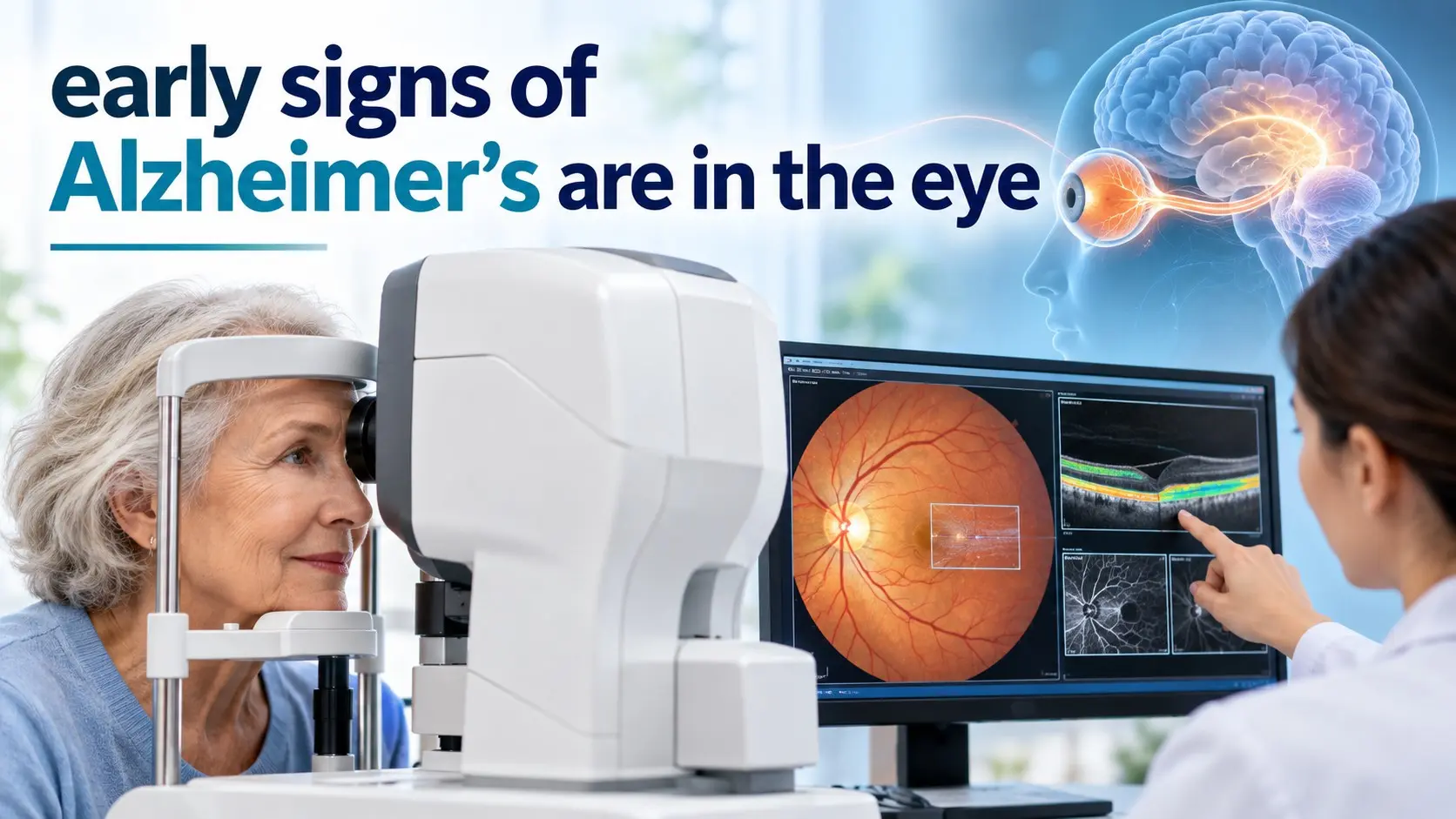
Early signs of Alzheimer's are in the eye is an exciting idea being studied by scientists, but it needs one important correction: eye changes may provide clues about Alzheimer’s biology, yet no routine eye examination can diagnose Alzheimer’s disease on its own.
The retina is part of the nervous system and is connected to the brain through the optic nerve. Researchers have found changes in retinal nerve layers, blood vessels, visual processing, pupil responses, and proteins such as amyloid in people along the Alzheimer’s disease spectrum.
That makes the eye a promising window into brain health.
It does not make every vision change a sign of dementia.
This article is for general information and education. It does not replace medical advice, diagnosis, or treatment.
New vision loss should not automatically be blamed on Alzheimer’s.
Sudden loss of vision, sudden double vision, a curtain or shadow across vision, many new flashes or floaters, severe eye pain, facial weakness, trouble speaking, or sudden confusion can be signs of an eye, brain, or circulation emergency.
Seek urgent medical care for sudden neurological or vision symptoms.
Table of Contents
- Quick Answer
- Can the eyes reveal Alzheimer’s?
- Can an eye test diagnose it?
- 9 Vital Research Clues
- Retinal nerve fibre changes
- Ganglion cell changes
- Retinal blood vessel changes
- Amyloid in retinal tissue
- Visual contrast changes
- Depth and spatial problems
- Pupil response changes
- Macular and choroidal findings
- Combined biomarkers
- OCT and OCT Angiography
- Vision Symptoms Versus Eye Biomarkers
- How Alzheimer’s Is Diagnosed Today
- When to Seek Help
- Frequently Asked Questions
Early Signs of Alzheimer's Are in the Eye: Quick Answer
The phrase early signs of Alzheimer's are in the eye comes from research showing that the retina and visual system may change during Alzheimer’s disease.
Scientists are studying:
- retinal nerve fibre layer thickness
- retinal ganglion cells
- retinal blood flow
- amyloid beta deposits
- OCT angiography findings
- visual contrast sensitivity
- pupil responses
These findings may one day help doctors detect or monitor Alzheimer’s earlier.
For now, however, retinal scans remain an area of active research. They are not a replacement for memory assessment, neurological evaluation, brain imaging, blood biomarkers, cerebrospinal fluid testing, or other established diagnostic methods.
Why Are Scientists Looking at the Eye?
The retina is the thin layer of nerve tissue at the back of the eye.
It turns light into electrical signals and sends those signals to the brain through the optic nerve.
The retina and brain share several important features:
- Nerve cells
- Small blood vessels
- Immune cells
- Similar developmental origins
- Connections through the central nervous system
This means that some disease processes affecting the brain may also leave measurable changes in the retina.
Unlike the brain, the retina can be examined quickly and without surgery.
This is why retinal biomarkers are so attractive to researchers.
9 Vital Eye Clues Researchers Are Studying
1. Retinal Nerve Fibre Layer Changes
The retinal nerve fibre layer contains nerve fibres that help carry visual information toward the optic nerve.
Several studies have reported thinning of this layer in people with Alzheimer’s disease or mild cognitive impairment.
This sounds promising, but the finding is not specific to Alzheimer’s.
Retinal nerve fibre thinning can also occur with:
- Glaucoma
- Normal ageing
- Optic nerve disease
- Other neurological conditions
A 2025 systematic review found that retinal nerve layer changes remain promising as research biomarkers, but results between studies are still inconsistent.
2. Retinal Ganglion Cell Changes
Retinal ganglion cells collect visual signals and send them toward the brain.
Researchers have found possible changes in the ganglion cell layer in people with early or established Alzheimer’s disease.
Some studies show thinning.
Others show different patterns at very early stages, which may reflect inflammation or compensation before later nerve loss occurs.
This variation is one reason doctors cannot yet use a single retinal thickness number to diagnose Alzheimer’s.
3. Retinal Blood Vessel Changes
The retina contains a fine network of blood vessels that can be photographed and measured.
Researchers have studied:
- retinal vessel density
- small blood vessel branching
- vessel width
- blood flow
- the foveal avascular zone
Some studies report reduced vessel density or changes in vascular patterns in people with Alzheimer’s.
However, diabetes, high blood pressure, smoking, ageing, and eye disease can also alter retinal blood vessels.
4. Amyloid Beta in the Retina
Amyloid beta is one of the hallmark proteins involved in Alzheimer’s disease.
An NIA supported study of donated retina and brain tissue found more retinal amyloid deposits in people with mild cognitive impairment and Alzheimer’s disease than in cognitively unimpaired people.
The amount of retinal amyloid also tracked with the severity of Alzheimer’s changes in the brain.
This finding is important.
It does not mean a standard eye doctor can currently look into your eye and see Alzheimer’s plaques during a routine appointment.
5. Visual Contrast Sensitivity
Vision involves much more than reading small letters.
Contrast sensitivity is the ability to tell an object apart from its background.
NIA funded research has found links between poorer contrast sensitivity and higher levels of amyloid and tau in the brain.
This raises the possibility that subtle changes in visual processing could provide early clues.
Larger studies are still needed before contrast testing becomes an Alzheimer’s screening test.
6. Depth, Distance, and Visual Processing Problems
Alzheimer’s disease affects the brain as well as memory.
Brain regions used to understand visual information may become damaged.
A person may have trouble:
- Judging distances
- Finding objects
- Reading
- Recognising faces
- Understanding complex scenes
- Separating an object from its background
These problems may occur even when the eye itself is healthy.
They are problems with how the brain interprets what the eyes send to it.
7. Pupil Response Changes
Your pupil changes size in response to light, attention, and mental effort.
Researchers have studied whether pupil dilation during thinking tasks might provide information about Alzheimer’s risk.
An NIA funded study found that greater pupil dilation during cognitive testing was associated with higher genetic risk for Alzheimer’s in a group of middle aged adults.
This is interesting research, not a diagnostic pupil test available at ordinary eye appointments.
8. Macular and Choroidal Changes
The macula is responsible for sharp central vision.
The choroid is a blood rich layer behind the retina.
Research has examined whether their thickness changes during Alzheimer’s disease.
Some studies report thinner retinal or choroidal layers.
Other studies find little difference or even different patterns at earlier stages.
Age, eye disease, blood pressure, diabetes, scan equipment, and study design can all influence the results.
9. Combining Eye Findings With Other Biomarkers
This may be the most realistic future direction.
An eye scan may eventually become more useful when combined with:
- Blood biomarkers
- Cognitive testing
- Medical history
- Genetic risk
- Brain imaging
- Other biological markers
Rather than one eye measurement giving a yes or no answer, several pieces of information may be combined to estimate risk more accurately.
What Is OCT?
Optical coherence tomography, or OCT, is a fast imaging test that creates detailed cross section pictures of the retina.
It uses light rather than X rays.
Eye professionals already use OCT to assess conditions such as:
- Macular degeneration
- Glaucoma
- Diabetic eye disease
- Retinal swelling
- Other retinal conditions
Alzheimer’s researchers use OCT because it can measure tiny changes in retinal layers.
What Is OCT Angiography?
OCT angiography, often shortened to OCTA, maps blood flow in small retinal vessels.
Researchers are investigating whether Alzheimer’s causes patterns such as:
- Lower capillary density
- Changes in blood flow
- A larger foveal avascular zone
- Altered retinal vessel networks
These measurements remain research tools for Alzheimer’s detection rather than routine diagnostic criteria.
Can an OCT Scan Diagnose Alzheimer’s?
No.
An OCT scan can provide useful information about the retina and optic nerve.
It can also detect many eye diseases.
Current evidence does not support diagnosing Alzheimer’s from OCT alone.
A 2025 systematic review of 22 studies found retinal structural and blood vessel changes in early Alzheimer’s research, but the findings varied substantially between studies.
The authors concluded that OCT and OCTA remain promising candidate biomarkers rather than established stand alone diagnostic tests.
What the Research Really Says
| Finding | Research Status | Can It Diagnose Alzheimer’s Alone? |
|---|---|---|
| Retinal nerve thinning | Reported in several studies | No |
| Ganglion cell changes | Promising but inconsistent | No |
| Retinal blood vessel changes | Associated in some studies | No |
| Retinal amyloid | Seen in tissue research | No routine clinical test yet |
| Visual contrast changes | Early research association | No |
| Pupil response | Research stage | No |
Can a Regular Eye Exam Find Alzheimer’s?
No regular eye exam is currently approved as a stand alone Alzheimer’s diagnostic test.
A comprehensive eye examination can still be very valuable.
It can identify conditions that can affect vision and daily function, including:
- Cataracts
- Glaucoma
- Macular degeneration
- Diabetic retinopathy
- Refractive errors
These conditions become more common with age and can sometimes make cognitive symptoms look worse.
Read the NextFitLife Eye Health Complete Guide for a full overview of routine eye care and warning signs.
Eye Problems Versus Brain Processing Problems
A person can have difficulty seeing for two very different reasons.
The Eye Itself May Be Unhealthy
Examples include:
- Cataracts
- Macular degeneration
- Glaucoma
- Diabetic retinopathy
The Eye May Be Healthy but the Brain Misreads the Image
Dementia can affect brain regions that process visual information.
This can make it difficult to:
- Judge depth
- Recognise objects
- Recognise faces
- Find items in a busy scene
- Understand reflections
- Navigate familiar places
An eye examination can help separate eye disease from neurological visual problems.
Visual Problems That Can Occur With Alzheimer’s
Alzheimer’s disease can affect visual processing as brain changes progress.
Possible difficulties include:
- Judging distance
- Recognising objects
- Reading
- Finding objects
- Navigating spaces
- Understanding contrast
These problems are not proof of Alzheimer’s.
Cataracts, macular degeneration, glaucoma, stroke, medication effects, and ordinary eyesight problems can cause similar difficulties.
What Is Posterior Cortical Atrophy?
Posterior cortical atrophy, or PCA, is a less common neurological syndrome in which visual and spatial problems can appear early.
It is sometimes caused by Alzheimer’s disease pathology.
People may struggle with:
- Reading
- Judging distance
- Finding objects
- Recognising what they see
- Using stairs
- Driving
Because vision problems are prominent, people may initially visit an eye professional even though the main problem is in the brain.
Are Floaters an Early Sign of Alzheimer’s?
No evidence shows that ordinary eye floaters are a typical early sign of Alzheimer’s.
Floaters usually come from changes inside the eye.
A sudden shower of new floaters, especially with flashes or a dark curtain in your vision, can signal a retinal tear or detachment.
That requires urgent eye assessment.
Are Dry Eyes a Sign of Alzheimer’s?
Dry eye is common, especially with age, screens, medications, and tear gland changes.
It is not considered a specific early sign of Alzheimer’s disease.
Do not assume burning, grittiness, or watering means dementia.
Are Cataracts a Sign of Alzheimer’s?
No.
Cataracts are clouding of the natural lens of the eye.
They are common with ageing and are not themselves a diagnosis of Alzheimer’s.
However, untreated vision loss can make daily functioning and cognitive testing more difficult.
Is Macular Degeneration a Sign of Alzheimer’s?
No.
Age related macular degeneration is a separate eye disease affecting central vision.
Some eye diseases and dementia share risk factors such as ageing and vascular health, but one does not automatically mean the other is present.
Can Glaucoma Look Like Alzheimer’s Eye Changes?
Yes, this is one reason retinal biomarkers can be difficult to interpret.
Glaucoma can cause thinning of the retinal nerve fibre layer and damage to the optic nerve.
If researchers are trying to use retinal thinning as an Alzheimer’s clue, they must carefully account for glaucoma and other eye disease.
Can Diabetes Affect the Same Retinal Blood Vessels?
Yes.
Diabetes can damage retinal blood vessels and cause diabetic retinopathy.
High blood pressure can also affect them.
That makes retinal vascular biomarkers less specific unless researchers carefully control for these conditions.
Does Amyloid in the Retina Prove Alzheimer’s?
No.
Retinal amyloid is a major research topic.
Postmortem studies have found relationships between retinal amyloid and Alzheimer’s changes in the brain.
However, scientists still need reliable, standardised methods that can detect disease specific retinal amyloid in living patients and prove how accurately it predicts Alzheimer’s.
Why Research Results Sometimes Conflict
Not every study finds the same retinal changes.
Several reasons may explain this:
- Small study groups
- Different OCT machines
- Different scan methods
- Different Alzheimer’s definitions
- Different disease stages
- Age differences
- Diabetes
- High blood pressure
- Glaucoma
- Other retinal disease
This is why standardisation is essential before retinal tests can become routine Alzheimer’s diagnostics.
How Is Alzheimer’s Diagnosed Today?
Doctors do not rely on one symptom or one scan.
A full evaluation may include:
- Medical history
- Memory testing
- Attention and language testing
- Daily function assessment
- Medication review
- Blood tests
- Neurological examination
- Brain imaging
- Biomarker testing when appropriate
Brain MRI and CT
Brain imaging can help doctors look for other causes of cognitive problems.
These may include:
- Stroke
- Tumours
- Hydrocephalus
- Other structural brain changes
MRI can also show patterns of brain shrinkage that may support a diagnosis.
Amyloid and Tau PET Scans
PET imaging can detect biological changes associated with Alzheimer’s.
An amyloid PET scan looks for abnormal amyloid plaques.
A tau PET scan looks for abnormal tau accumulation.
These tests are more specific to Alzheimer’s biology than an ordinary retinal scan.
Cerebrospinal Fluid Testing
Cerebrospinal fluid surrounds the brain and spinal cord.
A lumbar puncture can allow measurement of biomarkers such as:
- Amyloid beta
- Total tau
- Phosphorylated tau
These biomarkers can help support an Alzheimer’s diagnosis in selected patients.
Blood Tests for Alzheimer’s
This area has advanced quickly.
Blood tests can now measure Alzheimer’s related proteins such as forms of phosphorylated tau and amyloid.
In 2026, NIA information notes that FDA cleared blood testing is available in the United States to help assess adults aged 55 and older who already have signs or symptoms that may be related to Alzheimer’s.
Blood testing is not a reason to screen healthy people without symptoms on your own.
Results need clinical interpretation.
Why Early Diagnosis Matters
An accurate diagnosis can help people:
- Understand what is causing symptoms
- Rule out treatable conditions
- Discuss treatment choices
- Plan future care
- Address driving and safety
- Take part in clinical trials
- Discuss finances and legal planning
Early assessment also matters because not every memory problem is Alzheimer’s.
Memory Problems Can Have Other Causes
Problems with memory or attention may come from:
- Depression
- Anxiety
- Sleep problems
- Medication side effects
- Vitamin B12 deficiency
- Thyroid disease
- Hearing loss
- Vision loss
- Stroke
- Other neurological conditions
Some of these causes can be treated.
What Early Symptoms Matter More Than an Eye Scan?
For everyday clinical care, changes in memory, thinking, behaviour, and function remain important reasons to seek assessment.
Examples include:
- Repeating the same questions
- Forgetting recent events often
- Getting lost in familiar places
- Difficulty managing money
- Struggling with familiar tasks
- Word finding problems
- New problems judging distances
- Noticeable changes in personality
Normal Ageing Versus Possible Dementia
| Situation | Common Ageing | Worth Discussing With a Doctor |
|---|---|---|
| Names | Forget briefly, then remember later | Frequently forget familiar people |
| Appointments | Occasionally forget one | Repeatedly forget despite reminders |
| Navigation | Need directions in a new area | Get lost in familiar places |
| Vision | Need stronger glasses | Have trouble understanding what is being seen |
| Daily tasks | Take a little longer | Cannot complete familiar tasks safely |
What Should You Do if You Notice Vision and Memory Changes?
Do not choose between an eye appointment and a medical appointment.
You may need both.
Book an Eye Examination
This can identify:
- Cataracts
- Glaucoma
- Macular disease
- Prescription changes
- Retinal disease
Speak With Your Doctor
Discuss:
- Memory changes
- Behaviour changes
- Difficulty with familiar tasks
- Navigation problems
- Visual processing problems
Bring a relative or trusted person if they have noticed changes too.
Can Protecting Your Eyes Protect Your Brain?
Eye and brain health overlap in several ways.
Good habits include:
- Managing blood pressure
- Managing diabetes
- Not smoking
- Staying physically active
- Getting vision problems corrected
- Eating a balanced diet
- Staying socially engaged
Alzheimer’s Society reports that untreated vision loss is associated with a higher risk of dementia.
That does not mean ordinary glasses prevent Alzheimer’s directly.
It does mean that maintaining useful vision is an important part of healthy ageing.
Can Eye Vitamins Prevent Alzheimer’s?
No vitamin has been proven to prevent Alzheimer’s simply by improving eye health.
Eye supplements such as AREDS2 are designed for selected people with age related macular degeneration.
They are not Alzheimer’s prevention supplements.
For evidence based eye nutrition information, read our Eye Health Complete Guide.
Can an Optometrist Tell if You Have Alzheimer’s?
An optometrist may notice unusual visual problems or retinal findings that deserve further assessment.
They can also identify ordinary eye disease that may be affecting function.
They cannot diagnose Alzheimer’s simply by looking into the eye during a standard examination.
Will Eye Scans Replace Brain Scans?
Not yet.
Retinal imaging has several advantages:
- It is fast
- It is noninvasive
- It is widely available
- It is less expensive than many brain scans
The challenge is specificity.
The same retinal change can happen because of ageing, glaucoma, diabetes, high blood pressure, or another neurological condition.
A future eye based Alzheimer’s test will need to separate these causes reliably.
What Is the Most Promising Future Approach?
The strongest future strategy may be a combination rather than one test.
For example:
- A retinal OCT scan
- A blood biomarker
- A short cognitive test
- Medical history
- Risk factors
An algorithm could combine these signals and decide who needs more specific testing.
This could make early evaluation faster and more accessible.
5 Things the Eye Research Does Not Mean
- It does not mean every retinal change is Alzheimer’s.
- It does not mean an optician can diagnose Alzheimer’s from a routine eye test.
- It does not mean eye symptoms appear before memory symptoms in every person.
- It does not mean OCT replaces PET, CSF, blood biomarkers, or cognitive assessment.
- It does not mean vision changes should be ignored because they might be dementia.
Eye Symptoms That Need Urgent Care
Seek urgent eye or medical assessment for:
- Sudden vision loss
- A curtain or dark shadow over vision
- Many new floaters
- Flashing lights
- Sudden double vision
- Severe eye pain
- Eye pain with nausea
- Vision changes with facial weakness
- Vision changes with trouble speaking
These symptoms have urgent causes that are much more important to rule out immediately than Alzheimer’s.
A Simple Brain and Eye Health Checklist
Keep Eye Exams Current
Correct vision problems and monitor diagnosed eye disease.
Control Blood Pressure
Healthy blood vessels support both the retina and brain.
Manage Diabetes
Diabetes can damage small vessels in the eye and elsewhere in the body.
Avoid Smoking
Smoking harms blood vessels and increases the risk of several eye and brain diseases.
Move Regularly
Physical activity supports cardiovascular and brain health.
Pay Attention to Cognitive Changes
Do not wait for a retinal test if memory or thinking problems are affecting daily life.
Common Myths About Alzheimer’s and the Eye
Myth 1: An Eye Doctor Can Already Diagnose Alzheimer’s From the Retina
False.
Retinal Alzheimer’s biomarkers remain an active research area.
Myth 2: Thin Retinal Nerves Mean Alzheimer’s
False.
Glaucoma, ageing, neurological disease, and other conditions can also cause thinning.
Myth 3: Amyloid Can Be Seen During a Normal Eye Exam
False.
Research on retinal amyloid uses specialised methods.
Myth 4: Every Vision Problem in an Older Adult Is Dementia
False.
Cataracts, glaucoma, macular degeneration, and ordinary refractive problems are common.
Myth 5: A Normal Eye Test Rules Out Alzheimer’s
False.
A normal standard eye examination cannot rule out Alzheimer’s disease.
Myth 6: Eye Scans Have No Value for Alzheimer’s Research
False.
Retinal imaging is one of the most promising noninvasive research areas, but it still needs stronger validation and standardisation.
Conclusion
The statement early signs of Alzheimer's are in the eye captures an exciting area of research, but it should not be read as a proven home or routine eye test for Alzheimer’s.
Researchers have found associations involving retinal nerve layers, ganglion cells, blood vessels, amyloid deposits, visual contrast, and pupil responses. Modern OCT and OCT angiography make these changes easier to study without surgery.
The limitation is that these findings are not yet specific or consistent enough to diagnose Alzheimer’s by themselves.
The most promising future may combine retinal imaging with blood biomarkers, cognitive testing, medical history, and other measures.
Your next step is practical: keep routine eye care up to date, but speak with a healthcare professional if you or someone close to you develops persistent memory, thinking, navigation, or visual processing problems. Sudden vision or neurological symptoms need urgent assessment.
References and Sources
- National Institute on Aging. Changes in the Retina Linked to Stages of Alzheimer’s Disease.
https://www.nia.nih.gov/news/changes-retina-linked-stages-alzheimers-disease - National Institute on Aging. Two Views on Alzheimer's Biomarkers: Eyeing Changes in Vision or Pupils.
https://www.nia.nih.gov/news/two-views-alzheimers-biomarkers-eyeing-changes-vision-or-pupils - National Institute on Aging. How Is Alzheimer's Disease Diagnosed?
https://www.nia.nih.gov/health/alzheimers-symptoms-and-diagnosis/how-alzheimers-disease-diagnosed - National Institute on Aging. How Biomarkers Help Diagnose Dementia.
https://www.nia.nih.gov/health/alzheimers-symptoms-and-diagnosis/how-biomarkers-help-diagnose-dementia - Retinal Biomarkers for Early Alzheimer's Detection: A Systematic Review of Optical Coherence Tomography Findings.
https://pmc.ncbi.nlm.nih.gov/articles/PMC12829385/ - Retinal Imaging Biomarkers of Alzheimer's Disease: A Systematic Review and Meta Analysis.
https://pmc.ncbi.nlm.nih.gov/articles/PMC10210353/ - Alzheimer's Society. Sight and Hearing Loss With Dementia.
https://www.alzheimers.org.uk/about-dementia/stages-and-symptoms/sight-hearing-loss
Internal Links + Hub
- Eye Health Complete Guide
- Eye Problems NHS
- Eye Cancer
- Itchy Eyes
- Eye Vitamins
- How to Build and Maintain a Healthy Brain
- Healthy Eating for Older Adults
About the Author
Adel Galal is the founder and editor of NextFitLife. He writes practical consumer health, eye health, nutrition, fitness, brain health, and healthy ageing content using information from recognized medical, public health, and academic sources.
I am not a dermatologist, doctor, neurologist, ophthalmologist, optometrist, geriatrician, pharmacist, or other licensed healthcare professional. This content does not replace professional medical advice. What I share comes from real life experience, extensive research, and consultation with healthcare providers. Always consult qualified medical professionals for diagnosis and treatment of any health condition.
Learn more about NextFitLife, the author, and the site's editorial approach on the About Us page.
Frequently Asked Questions
Are early signs of Alzheimer's in the eye?
Research suggests that Alzheimer’s disease may be associated with changes in the retina, retinal blood vessels, visual processing, pupil responses, and retinal proteins. These findings are promising biomarkers, but they are not yet enough to diagnose Alzheimer’s from an eye test alone.
Can an eye test detect Alzheimer's disease?
Not by itself. Routine eye examinations can find eye disease, but Alzheimer’s diagnosis still requires clinical assessment and may include cognitive testing, brain imaging, blood biomarkers, cerebrospinal fluid testing, or other specialist evaluation.
Can OCT detect early Alzheimer's?
OCT can identify retinal structural changes associated with Alzheimer’s in research studies, but current evidence is too variable and nonspecific for OCT to diagnose Alzheimer’s alone.
What retinal changes are linked with Alzheimer's?
Studies have reported retinal nerve fibre changes, ganglion cell changes, retinal blood vessel differences, macular changes, and possible retinal amyloid deposits.
Can Alzheimer's cause vision problems?
Yes. Alzheimer’s can affect brain regions that process visual information, which may cause difficulty judging distance, recognising objects, reading, navigating spaces, or understanding what is being seen.
Are floaters a sign of Alzheimer's?
No. Ordinary floaters are not considered a typical early sign of Alzheimer’s. Sudden new floaters with flashes or a curtain over vision can signal a retinal tear or detachment and need urgent assessment.
Are dry eyes a sign of Alzheimer's?
No. Dry eye is common and is usually related to tear film problems, ageing, medicines, screens, or environmental factors rather than Alzheimer’s disease.
Can retinal amyloid diagnose Alzheimer's?
Not currently in routine care. Retinal amyloid has been found in research studies, including postmortem tissue, but reliable standardised clinical retinal amyloid testing is still being developed.
Can an optometrist diagnose Alzheimer's?
No. An optometrist can identify eye conditions and may notice visual problems that deserve further investigation, but Alzheimer’s diagnosis requires medical and cognitive assessment.
What is the most accurate way to diagnose Alzheimer's?
Diagnosis uses a combination of medical history, cognitive testing, neurological assessment, laboratory tests, and sometimes biomarkers such as amyloid or tau PET, cerebrospinal fluid markers, or validated blood biomarkers.
What should I do if memory and vision are both changing?
Arrange both an eye examination and a medical assessment. Eye disease and neurological problems can overlap, and treating vision loss may improve daily function even when cognitive problems are also present.
When are vision changes an emergency?
Seek urgent help for sudden vision loss, sudden double vision, a curtain over vision, many new flashes or floaters, severe eye pain, or vision changes with weakness, facial droop, or trouble speaking.

Health & wellness writer with 30+ years of experience in nutrition, fitness, and healthy aging. Founder of NextFitLife.com — evidence-based health guidance.