
First published: May 7, 2023
Last updated: September 30, 2026
Written by: Adel Galal, Founder and Editor of NextFitLife
Medical review status: This article has not been medically reviewed.
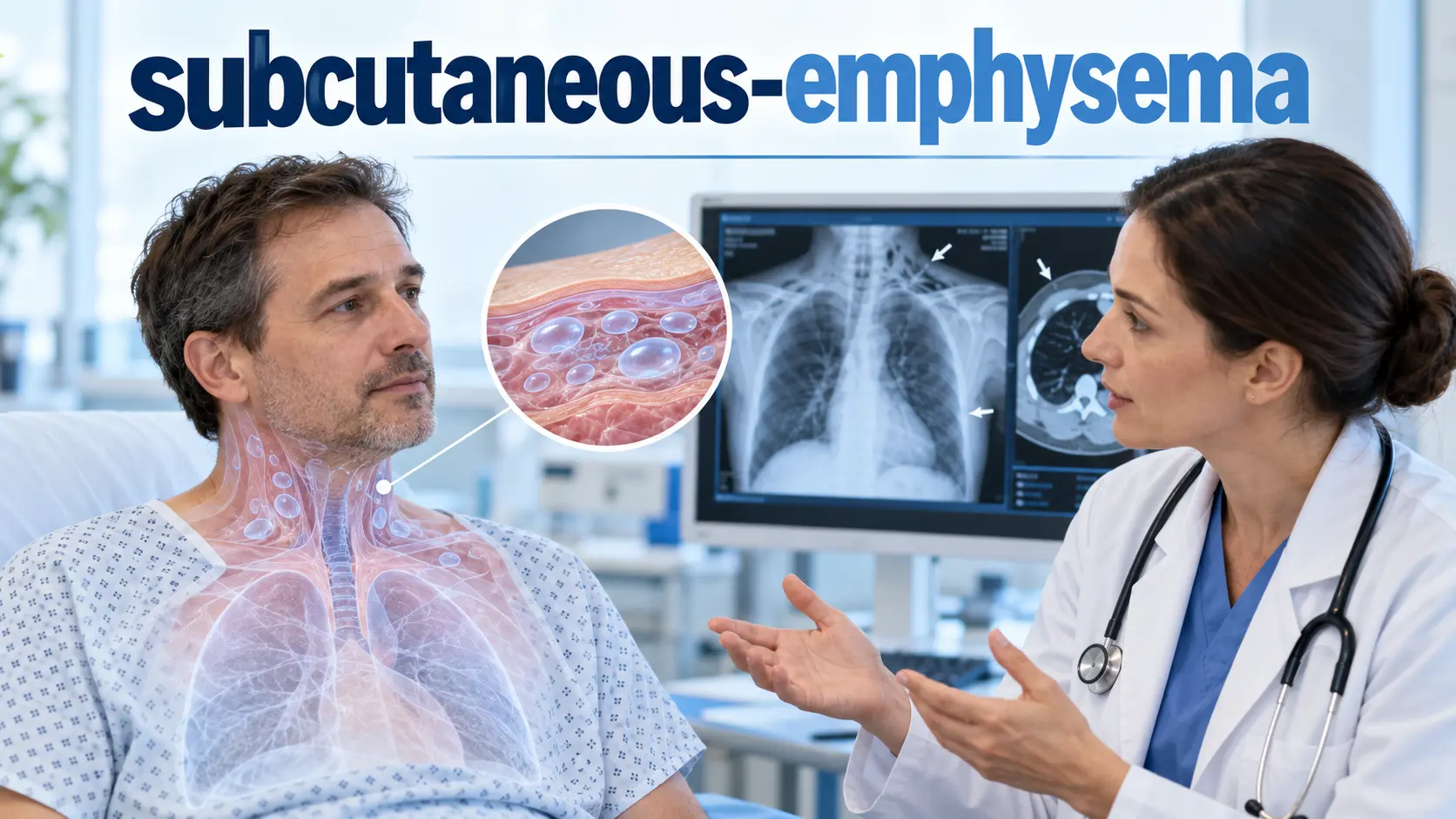
Subcutaneous Emphysema means air or gas has moved into tissue beneath the skin. It often causes swelling and a strange crackling feeling called crepitus.
The trapped air itself may be small and temporary, but the cause matters. A collapsed lung, chest injury, airway tear, medical procedure, severe infection, or another air leak can sit underneath the visible swelling.
Quick Answer
Subcutaneous emphysema is air trapped beneath the skin, most often around the chest, neck, or face. Common causes include chest trauma, pneumothorax, surgery, chest drains, airway procedures, ventilation, and some infections. Treatment focuses on stopping the source of the air leak. Breathing trouble or rapidly spreading swelling needs urgent care.
Get Emergency Help for These Symptoms
Seek emergency medical care if swelling under the skin occurs with severe or worsening breathlessness, sudden chest pain, rapidly increasing neck or facial swelling, trouble swallowing, noisy breathing, blue or grey lips, fainting, confusion, or collapse.
Subcutaneous emphysema after major chest or neck trauma can point to a serious hidden injury. Do not try to puncture, squeeze, massage, or drain the air yourself.
Medical notice: This content is for general educational purposes and does not replace professional medical advice, diagnosis, or treatment. Consult an appropriately qualified healthcare professional for personal medical concerns. This article explains published guidance and does not provide a personal clinical assessment. Always consult qualified medical professionals for diagnosis and treatment of any health condition.
Table of Contents
- What Subcutaneous Emphysema Means
- How It Differs From Lung Emphysema
- 9 Critical Facts
- Common Causes
- Symptoms and Crepitus
- How Air Spreads Through the Body
- Diagnosis and Imaging
- Treatment Options
- Chest Drains and Surgical Emphysema
- Serious Complications
- Recovery
- What Not to Do at Home
- When to Seek Emergency Care
- References and Sources
- Related NextFitLife Guides
- About the Author
- Frequently Asked Questions
What Is Subcutaneous Emphysema?
Subcutaneous Emphysema happens when air enters the layer of tissue beneath the skin.
It most often appears around the chest or neck, but air can also spread to the face, abdomen, arms, or other areas.
The skin may look puffy or swollen. When the area is touched, it can feel as though tiny bubbles are moving beneath the fingers.
That crackling feeling is called crepitus.
MedlinePlus also uses the terms subcutaneous air, tissue emphysema, crepitus, and surgical emphysema for this finding.
Is Subcutaneous Emphysema the Same as Lung Emphysema?
No.
The names sound similar, but the two conditions are very different.
| Condition | Where the Problem Is | What Happens |
|---|---|---|
| Subcutaneous emphysema | Beneath the skin | Air escapes into soft tissue |
| Lung emphysema | Inside the lungs | The walls of lung air sacs become damaged |
If you meant chronic lung emphysema linked with COPD, read our Emphysema NHS guide.
9 Critical Facts About Subcutaneous Emphysema
1. It Is a Sign, Not Always the Main Disease
The air under the skin is often a clue to another problem.
The most useful question is not only, “How do we remove the air?” It is, “Where is the air coming from?”
A person may have an injured lung, a pneumothorax, a damaged airway, a chest drain problem, a tear in the digestive tract, or another source.
Finding and treating that source comes first.
2. The Crackling Feeling Has a Name
The classic physical sign is crepitus.
Pressing gently over the swollen area can produce a crackling or popping feeling because tiny pockets of air move through the tissue.
Some people compare the sensation with bubble wrap or crisp cereal beneath the skin.
You do not need to keep pressing the area once you notice this sign.
3. The Air Can Travel Far From Where It Started
Air follows spaces between layers of tissue.
An air leak that begins in the chest can move upward into the neck and face or downward through the chest wall and abdomen.
This explains why the swelling may appear some distance from the original injury or procedure.
4. A Collapsed Lung Is an Important Cause
A pneumothorax happens when air collects between a lung and the chest wall.
That air can move into nearby soft tissue and cause subcutaneous emphysema.
A pneumothorax can occur after trauma, a procedure, lung disease, mechanical ventilation, or sometimes without an obvious trigger.
Sudden chest pain and breathlessness need prompt medical assessment.
5. Surgery Is Only One Cause
When subcutaneous emphysema develops after an operation, it is often called surgical emphysema.
It can follow chest surgery, laparoscopy, chest drain insertion, intubation, bronchoscopy, dental procedures, or other procedures that allow air to enter tissue.
If your symptoms started after an operation or chest drain, see our focused Surgical Emphysema guide.
6. Infection Can Also Produce Gas Under the Skin
Most subcutaneous emphysema is related to escape air rather than infection.
Still, some serious bacterial infections can make gas inside soft tissue.
Gas gangrene and some necrotising infections can cause severe pain, rapid swelling, fever, dark skin changes, blisters, foul drainage, low blood pressure, or rapid illness.
This pattern is a medical emergency.
7. X Ray and CT Can Help Find the Source
Doctors can often suspect the condition from the history, swelling, and crepitus.
Imaging helps answer the more important question: where did the air come from?
A chest X ray may show subcutaneous air, pneumothorax, or pneumomediastinum.
A CT scan gives more detail and can show how far the air has spread.
8. Mild and Severe Cases Are Treated Differently
A small amount of air may slowly disappear once the leak has stopped.
A severe case may require treatment of a pneumothorax, adjustment of a chest drain, repair of an airway or digestive tract injury, antibiotics for infection, or drainage of the subcutaneous air.
There is no single treatment that fits every case.
9. Breathing and Airway Symptoms Change the Urgency
Swelling alone may be uncomfortable but stable.
Rapid spread toward the neck or face is more concerning because large amounts of air can place pressure on the airway and surrounding structures.
Difficulty breathing, swallowing, or speaking changes the situation from simple observation to urgent assessment.
What Causes Subcutaneous Emphysema?
The cause usually falls into a few broad groups.
Chest Trauma
Blunt or penetrating trauma can damage the lung, chest wall, airway, or ribs.
A rib fracture may injure lung tissue and allow air to escape.
Subcutaneous emphysema after major trauma should not be treated as a harmless skin problem because it can signal deeper chest or airway injury.
Pneumothorax
A collapsed lung is one of the best known causes.
Air escapes from the lung and may enter both the pleural space and the tissue beneath the skin.
Pneumomediastinum
Pneumomediastinum means air has entered the central part of the chest between the lungs.
That air can then travel upward into the neck and soft tissues.
Possible triggers include intense coughing, vomiting, asthma, trauma, procedures, and lung injury.
Airway Injury
A tear in the larynx, trachea, or bronchial tubes can leak air into surrounding tissues.
This can happen after trauma or, rarely, after intubation or another airway procedure.
Voice changes, neck swelling, breathing trouble, or coughing blood after an airway injury need urgent attention.
Oesophageal or Digestive Tract Injury
A tear in the oesophagus can also release air into the chest and neck.
A rare emergency called Boerhaave syndrome can occur after forceful vomiting. Severe chest pain, swallowing pain, breathing difficulty, and subcutaneous emphysema can occur.
Medical Procedures
Possible procedure related causes include:
- Chest drain placement
- Thoracic surgery
- Bronchoscopy
- Intubation
- Mechanical ventilation
- Endoscopy
- Central venous line procedures
- Laparoscopic surgery
- Some dental procedures
Positive Pressure Ventilation
Positive pressure ventilation pushes air into the lungs.
If fragile or injured lung tissue leaks, air can escape into the chest and soft tissues.
Pressure Injury and Diving
Rapid pressure changes can injure the lungs during diving.
Pulmonary barotrauma can lead to pneumothorax, pneumomediastinum, subcutaneous emphysema, or other serious pressure related problems.
Gas Producing Infection
Some severe infections produce gas directly inside soft tissue.
These are very different from a small air leak after a procedure.
Severe pain, fever, fast progression, skin colour change, blisters, or signs of shock require emergency care.
What Are the Symptoms?
The most common visible symptom is swelling.
The most characteristic physical finding is crepitus.
Other symptoms depend on the amount of air and the underlying cause.
- Chest swelling
- Neck swelling
- Facial swelling
- Puffy eyelids
- Crackling beneath the skin
- Chest discomfort
- Neck pain
- Sore throat
- Voice change
- Difficulty swallowing
- Shortness of breath
Breathlessness may come from the condition underneath rather than from the skin swelling itself.
Why Can the Face and Neck Swell?
Soft tissue layers are connected.
Once air enters one area, pressure can push it along those tissue planes.
Air from the chest can rise into the neck and face. Severe cases may cause marked eyelid swelling or change the voice.
Rapid progression toward the airway needs urgent medical assessment.
How Is Subcutaneous Emphysema Diagnosed?
Doctors begin with the story and the examination.
History
Useful details include recent:
- Chest injury
- Surgery
- Chest drain
- Intubation
- Mechanical ventilation
- Dental treatment
- Endoscopy
- Forceful vomiting
- Severe coughing
- Diving
- Signs of infection
Physical Examination
The healthcare team checks swelling, breathing, oxygen level, pulse, blood pressure, chest movement, breath sounds, neck, and other affected areas.
Crepitus is an important clue.
Chest X Ray
An X ray can show air in the soft tissue and may reveal pneumothorax or pneumomediastinum.
CT Scan
A CT scan can show the exact spread of air in much more detail.
It can also help doctors find damage to the lungs, airway, mediastinum, chest wall, or abdomen.
Ultrasound
Bedside ultrasound may help identify a pneumothorax in an emergency setting.
Subcutaneous air itself can sometimes make ultrasound harder because air blocks the sound waves.
Bronchoscopy or Other Specialist Tests
Bronchoscopy may be used when an airway injury is suspected.
An oesophageal tear or digestive tract injury may need different imaging or endoscopic assessment.
How Is Subcutaneous Emphysema Treated?
The treatment rule is simple:
Treat the source of the air first.
If the leak continues, removing only the visible air does not solve the problem.
Observation
A small stable collection may be monitored when the patient is well and serious causes have been excluded or treated.
The body can gradually absorb the trapped air.
Oxygen
Supplemental oxygen may be used in hospital for selected patients.
It can support people with low oxygen levels and, in some circumstances, may help trapped air reabsorb more quickly.
Oxygen should be prescribed and monitored by healthcare professionals.
Treating Pneumothorax
If a collapsed lung is causing the leak, the pneumothorax needs treatment.
Some people require a chest drain so air can leave the space around the lung.
Checking a Chest Drain
The British Thoracic Society notes that problematic surgical emphysema after chest drainage can occur when there is a large air leak, poor drain position, migration of drain holes into the subcutaneous tissue, or blockage.
The drain should be assessed by trained healthcare professionals.
Never reposition, clamp, or remove a chest drain yourself.
Treating an Airway Tear
A significant tracheal or bronchial injury may need specialist respiratory or thoracic surgical care.
Treating Oesophageal Injury
An oesophageal rupture is a medical emergency and can lead to severe infection in the chest.
Urgent specialist treatment is required.
Treating Infection
A serious soft tissue infection may need urgent antibiotics and surgery to remove damaged tissue.
Does the Air Ever Need to Be Drained From Under the Skin?
Yes, but only in selected severe cases.
Small collections usually do not need to be punctured or drained directly.
Extensive subcutaneous emphysema may need treatment when swelling becomes severe, painful, rapidly progressive, closes the eyes, or contributes to airway pressure.
Hospital techniques can include:
- Subcutaneous catheters
- Small drainage tubes
- Release incisions
- Negative pressure wound therapy in selected cases
These methods are performed by trained clinicians.
The evidence for severe decompression methods comes largely from case reports, case series, and observational experience rather than one standard procedure for every patient.
What Complications Can Occur?
| Complication | Why It Matters | Possible Warning Signs |
|---|---|---|
| Pneumothorax | A lung can partly or fully collapse | Chest pain and breathlessness |
| Tension pneumothorax | Pressure can affect breathing and circulation | Severe breathlessness, collapse, confusion |
| Pneumomediastinum | Air enters the central chest | Chest pain, neck pain, swallowing trouble |
| Airway compression | Large amounts of air can narrow the airway | Noisy breathing, voice change, neck swelling |
| Circulation problems | Extreme tissue pressure can reduce blood flow | Severe swelling and rapid deterioration |
| Severe infection | Gas producing infections can cause tissue death and sepsis | Severe pain, fever, skin changes, rapid illness |
Why Is Tension Pneumothorax So Serious?
A tension pneumothorax is different from a small stable air leak.
Pressure builds inside the chest. The affected lung becomes compressed, and the pressure can interfere with blood returning to the heart.
A person can deteriorate quickly.
Severe breathing difficulty, collapse, low blood pressure, confusion, or extreme distress after chest injury or a known pneumothorax needs emergency care.
How Long Does Subcutaneous Emphysema Last?
Recovery depends on what caused it and whether the leak has stopped.
Cleveland Clinic notes that many treated cases improve within about 10 to 14 days.
That is not a deadline.
A small collection can improve sooner. A large collection with an ongoing pneumothorax, major surgery, trauma, or airway injury can take longer.
Visible swelling is only part of recovery. The healthcare team also needs to know that the original cause is improving.
Can Subcutaneous Emphysema Go Away on Its Own?
Small amounts of trapped air can be absorbed naturally once the source stops.
That does not mean unexplained subcutaneous emphysema should simply be watched at home.
The same skin finding can occur with harmless postoperative air or with a serious pneumothorax, airway tear, digestive tract injury, or infection.
A healthcare professional should decide whether observation is safe.
What Should You Watch After Surgery or a Procedure?
Contact the treating team if you develop:
- New swelling
- Crackling beneath the skin
- Swelling that spreads
- New shortness of breath
- Chest pain
- Voice change
- Difficulty swallowing
- Fever
- Worsening pain
Do not assume all swelling after surgery is normal.
If the condition developed after an operation, chest drain, bronchoscopy, or other procedure, our Surgical Emphysema guide explains the procedure related causes in more detail.
What Should You Not Do at Home?
- Do not puncture the swollen skin.
- Do not cut the skin to release air.
- Do not forcefully massage the swelling.
- Do not move a chest drain.
- Do not clamp chest drain tubing yourself.
- Do not borrow oxygen equipment.
- Do not rely on one normal pulse oximeter reading when symptoms are severe.
- Do not delay assessment after significant trauma.
The skin swelling does not show you where the leak started.
When Is Subcutaneous Emphysema an Emergency?
Seek emergency care when you have:
- Severe or worsening difficulty breathing
- Sudden severe chest pain
- Rapid neck or facial swelling
- Noisy breathing
- Difficulty speaking
- Difficulty swallowing
- Blue or grey lips or skin
- Fainting or collapse
- Confusion or unusual drowsiness
- Coughing blood after an injury
- Severe pain, fever, or skin colour change suggesting infection
After major chest or neck trauma, new subcutaneous emphysema should receive urgent assessment even if the person initially looks fairly well.
Questions to Ask Your Healthcare Team
- Where is the air coming from?
- Do I have a pneumothorax?
- Is there air in the mediastinum?
- Do I need a CT scan?
- Is my airway safe?
- Is my chest drain working correctly?
- Could an infection be causing the gas?
- Does the air need direct drainage?
- What symptoms mean I should return urgently?
- When should the swelling begin to improve?
Conclusion
Subcutaneous Emphysema is air beneath the skin. The swelling and crackling can look dramatic, but the bigger concern is where the air came from.
Some cases settle once a small air leak stops. Others are linked with pneumothorax, airway injury, pneumomediastinum, serious infection, or a complication of surgery or chest drainage.
If you notice new crackling swelling, get it assessed. If it spreads quickly or comes with chest pain, severe breathlessness, trouble swallowing, fainting, or confusion, seek emergency care.
References and Sources
These medical and professional sources support the definition, causes, complications, diagnosis, chest drain guidance, and treatment information in this article.
- MedlinePlus, U.S. National Library of Medicine.
Subcutaneous Emphysema.
Reviewed July 12, 2024. Covers definition, crepitus, trauma, pneumothorax, airway and oesophageal injury, procedures, infection, imaging, and emergency evaluation.
https://medlineplus.gov/ency/article/003286.htm - Cleveland Clinic.
Subcutaneous Emphysema: What It Is, Causes and Treatment.
Medically reviewed. Updated March 27, 2025. Covers symptoms, complications, diagnosis, treatment, decompression, and recovery.
https://my.clevelandclinic.org/health/diseases/subcutaneous-emphysema - Kukuruza K, Aboeed A.
Subcutaneous Emphysema.
StatPearls. NCBI Bookshelf. StatPearls Publishing 2026.
Explains spread through tissue planes, pneumothorax, pneumomediastinum, severe expansion, respiratory compromise, and evaluation.
https://www.ncbi.nlm.nih.gov/books/NBK542192/ - British Thoracic Society.
British Thoracic Society Clinical Statement on Pleural Procedures.
Thorax. Includes troubleshooting guidance for surgical or subcutaneous emphysema after chest drain placement and identifies drain blockage, poor position, migration, and large air leaks as important causes.
https://thorax.bmj.com/content/78/Suppl_3/s43 - Lim CW, Sia LC.
Management of Severe Subcutaneous Emphysema With a Subcutaneous Cannula: A Life Saving Approach.
Cureus. 2024;16(12):e75865.
Open access clinical report describing decompression in severe subcutaneous emphysema.
https://pmc.ncbi.nlm.nih.gov/articles/PMC11736977/
Related NextFitLife Guides and Lung Health Hub
This article is the broad NextFitLife guide to subcutaneous emphysema. Use the related pages below for narrower questions about procedure related air leaks, chronic lung emphysema, and respiratory health.
- NextFitLife Lung and Respiratory Health Hub
- Surgical Emphysema: Risks, Causes, and Urgent Care
- Emphysema NHS: Causes, Tests, and Treatment
- Left Side Chest Pain: Causes and Warning Signs
About the Author
Adel Galal is the founder and editor of NextFitLife. He writes consumer health, fitness, nutrition, sleep, and wellness content using recognized medical, public health, and academic sources.
This content is for general educational purposes and does not replace professional medical advice, diagnosis, or treatment. Consult an appropriately qualified healthcare professional for personal medical concerns. This article explains published guidance and does not provide a personal clinical assessment. Always consult qualified medical professionals for diagnosis and treatment of any health condition.
Learn more about Adel, NextFitLife, and the site's editorial approach on the About Us page.
Frequently Asked Questions
What is subcutaneous emphysema?
Subcutaneous emphysema is air or gas trapped in tissue beneath the skin. It most often affects the chest, neck, or face and can cause swelling and a crackling feeling called crepitus.
Is subcutaneous emphysema the same as lung emphysema?
No. Lung emphysema damages the air sacs inside the lungs. Subcutaneous emphysema means air has escaped into tissue beneath the skin.
What causes subcutaneous emphysema?
Causes include chest trauma, pneumothorax, pneumomediastinum, airway injury, oesophageal injury, surgery, chest drains, intubation, mechanical ventilation, dental procedures, diving injuries, and some gas producing infections.
What does subcutaneous emphysema feel like?
The skin may look swollen and feel crackly or bubbly when touched. The crackling sensation is called crepitus.
Is subcutaneous emphysema dangerous?
It can be mild, but it can also be a sign of a serious underlying problem such as pneumothorax, airway injury, pneumomediastinum, or severe infection. Rapid spread or breathing symptoms need urgent care.
Can subcutaneous emphysema go away on its own?
Small amounts of air can be absorbed after the air leak stops. A healthcare professional should still identify the cause and decide whether observation is safe.
How is subcutaneous emphysema treated?
Treatment focuses on the source of the air. Options can include observation, oxygen, treatment of a pneumothorax, chest drainage, correction of a chest drain problem, repair of an airway or digestive tract injury, antibiotics, or direct decompression in severe cases.
How long does subcutaneous emphysema last?
The timeline varies. Cleveland Clinic notes that many treated cases improve within about 10 to 14 days, but recovery can be longer when the air collection is large or the underlying leak continues.
What is the difference between surgical emphysema and subcutaneous emphysema?
Surgical emphysema is a name often used for subcutaneous emphysema that occurs after surgery, chest drainage, or another medical procedure. Subcutaneous emphysema is the broader medical term.
When should I seek emergency help?
Seek emergency care for severe breathlessness, sudden chest pain, rapidly spreading neck or facial swelling, trouble speaking or swallowing, blue or grey skin, fainting, confusion, or collapse.

Health & wellness writer with 30+ years of experience in nutrition, fitness, and healthy aging. Founder of NextFitLife.com — evidence-based health guidance.